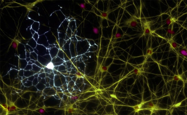
When we refer to the human brain, we generally think that it is made up of neurons that determine our thinking and intelligence. Well this is true only in a small percentage.
The human brain is made up of more than 80.000 million neurons, but this figure comes to represent only 15% of the total cells of the organs that compose it.
Cells and their function within the human body
The other 85% is made up of other microscopic cells called glial cells, responsible for forming a substance called glia that extends to all corners of the nervous system.
Glial cells are responsible for helping neurons in their process of transmitting electrochemical impulses through the nervous system. Glial cells are responsible for providing nutrients, maintain the structure or accelerate neural conduction itself, repair damage and provide energy to neurons.
Among these many glial cells found in the brain, the so-called oligodendrocytes for its ability to form the protective myelin sheaths of the axons of the central nervous system.
- Myelin it is a lipoprotein that allows the expansion of the action potential in time and distance. They protect the axon from the electrical impulse by making its path and prevents its dispersion through the neuronal membrane.
- Oligodendrocytes, Schwann cells, astrocytes, and microglia are the four most important classes of glial cells.
Schwann cells
They are the only ones found in the nerves that run throughout the body. (Peripheral nervous system). They are a kind of microscopic pearl-like sheaths composed of myelin
They are able to segregate the "Nerve growth factor" (NCF), a molecule that stimulates neuronal growth during development.
Schwann cells are responsible for the formation of myelin in the peripheral nervous system. Schwann cells are coiled around a single axon through its cytoplasm.
Astrocytes
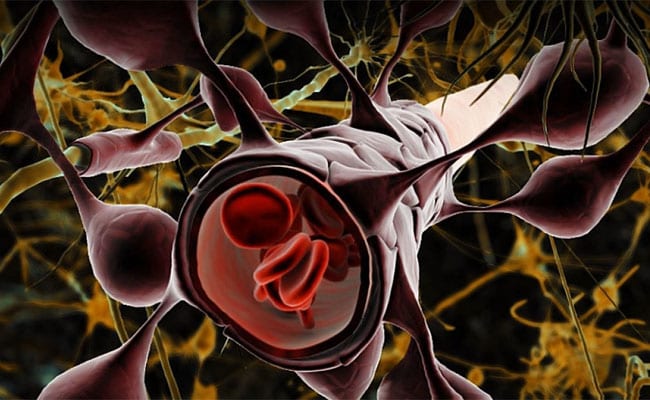
They are cells that are close to neurons, they are stellate in appearance, larger in size compared to neurons, they are found in the system central nervous (CNS) and by the optic nerve.
Astrocytes They are a kind of soldiers who are members of the Blood-brain Barrier (BBB), which is a protective membrane of the CNS whose function is to prevent blood from flowing directly into it.
Astrocytes are responsible for filtering what may or may not happen to the CNS. They allow the entry of oxygen and glucose; neurons nourishment.
microglia
It is the group of cells that make up the basis of the brain's immune system. Because the Blood-brain Barrier does not allow the free passage of the cells of the immune system, the brain has its own defensive system and these cells are its protective soldiers.
The basic function of these cells is to defend and repair the brain from injury caused by invading microorganisms, cell debris, and disease.
They constantly scan the CNS for damaged plaques, neurons and infectious agents. They are sensitive to the environment and are able to detect the smallest changes in the biological composition of brain tissue. Cells scan the CNS to locate and neutralize any plaque, deoxyribonucleic acid (DNA) fragments, neuronal tangles, dead cells, damaged cells, and foreign materials. They can be considered the housewives of the brain by cleaning the cellular debris.
Oligodendrocytes
It is a type of cell that is responsible for forming the myelin sheaths that surround the axons of the central nervous system. They are located only in the brain and in the bone marrow (CNS). They have many processes that wrap around the axons of various neurons.
The myelin sheaths created around the axons of neurons are intended to isolate them and increase the speed of transmission of electrochemical impulses.
This myelination is formed in the medulla spinal cord around week 16 of intrauterine life and progresses after birth until practically all nerve fibers are myelinated by the time the child begins to walk. Even in human adulthood, oligodendrocytes continue to reproduce from stem cells.
Types of oligodendrocytes
Oligodendrocytes can be classified mainly by their functions, although structurally and molecularly they are very similar. There are two main types: interfascicular and satellite.
- The interfascicular oligodendrocytes, are responsible for the formation of myelin sheaths, they make up part of the white matter of the brain.
- The satellite oligodendrocytes, they make up part of the gray matter, they do not produce myelin, they do not adhere to neurons, nor do they perform an isolation function. Its functions are unknown.
Features
Since it is not known exactly what the functions of satellite oligodendrocytes are, we will only go into the description of the functions of interfascicular ones.
Neural transmission acceleration
The speed of the action potentials increases when the axons have been myelinated. El correct system operation hormonal and muscular is favored before an adequate rhythm of neural conduction. Intelligence is also favored by the action of these cells on neurons.
Cell membrane isolation
Isolation of neuronal axons from the outer environment of cells prevents ion leakage through the cell membrane.
Structuring the nervous system
Since neurons are not capable of perform their function alone, glial cells, especially interfascicular oligodendrocytes, are responsible for supporting the network structure of neurons.
Support for the development of neurons
Oligodendrocytes are protein producers that, in their interaction with neurons, keep them active, thus preventing programmed cell death.
Extracellular fluid homeostasis
Despite that satellite oligodendrocytes have no clear function, are important to maintain the homeostatic balance of the external environment of the neurons close to them.
Diseases associated with myelin
Miller Fisher syndrome
It is a variant of Guillain-Barré syndrome, an autoimmune disease characterized by the production of antibodies against myelin in neurons of the peripheral nervous system.
Signal conduction is lost between the body and CNS, leading to potentially severe muscle paralysis. The functioning of the sense organs is also lost.
Symptoms associated with this disease are ophthalmology, ataxia and areflexia. If it is attended on time, it has good expectations of long-term improvement
Charcot – Marie – Tooth disease, or CMT
It is a hereditary disease that affects the peripheral nerves, it is known as peripheral neuropathy. It causes peripheral nerve damage, the most common cause is diabetes.
Multiple sclerosis
A disease of the nervous system that blocks or slows communication between the brain and the body. This occurs when the myelin sheath that protects the nerve cells is injured, affects the brain and marrow spinal.
The most frequent symptoms are due to loss of balance, involuntary muscle movements, movement problems, coordination difficulties, tremor, weakness, constipation or intestinal disorders.
Amyotrophic lateral sclerosis (ALS)
It progressively attacks motor neurons, which control involuntary muscles. They are characterized by gradual degeneration to the neuronal and organism death.
Baló's disease or Baló's concentric sclerosis
It generally affects children and rarely adults. It consists of the loss of myelin in the brain. It is rare and its It causes progressive paralysis, involuntary movements of the muscles among other neurological problems.
Leuko-dystrophies
It consists of alterations of the vision and motor system. It is caused by the destruction of myelin by enzymatic defects in the formation or maintenance of myelin or by processes of infectious, autoimmune, inflammatory or toxic vascular origin.